

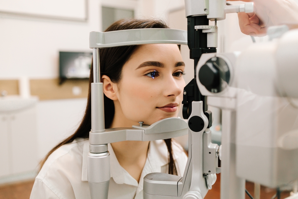
A field of vision exam catches vision loss most people never notice happening. Peripheral vision can fade for months before it affects daily tasks. This guide explains what the test checks, which conditions it catches, and how different testing methods compare. You’ll also learn when doctors recommend one and what the results usually mean.
Key Takeaways
- Visual acuity tests measure sharpness at the center of your vision, but optic nerve damage and visual loss can build silently in the surrounding field where acuity tests don’t reach.
- Conditions like retinitis pigmentosa, Diabetic retinopathy, brain tumors, and Optic Neuritis each produce specific patterns of peripheral vision loss that only a visual field test can map.
- From the dome-shaped Humphrey Field Analyzer to manual Goldmann perimetry, the type of test used depends on what the clinician is looking for and what the patient can tolerate.
- Catching side vision loss early opens the door to treatment options that can prevent visual disability before it becomes permanent.
- Get answers to the most common questions patients ask about visual field testing, including what to expect and who needs one.
Why Optic Nerve and Central Vision Health Depend on More Than Visual Acuity
Visual acuity testing only checks how clearly you see straight ahead. It does not measure what happens at the edges of your vision, where damage often starts. A visual field test fills that gap by mapping your entire range of sight, not just the center. Your eye doctor relies on this wider view to catch problems a standard eye chart misses.
Your optic nerve carries visual signals from the retina to the brain. Damage to this nerve, common in glaucoma, often shrinks the outer field first while central vision stays sharp. That is why the loss can go unnoticed for a long time. Patients rarely feel it happening until a large portion of their side vision is already gone.
Certain groups face a higher risk of unnoticed vision loss. Older adults, people with diabetes, and those with a family history of glaucoma should ask about visual field testing at their next visit. African American and Hispanic patients also face elevated glaucoma risk and may benefit from earlier screening.
Even without symptoms, these risk factors alone can justify more frequent testing. Talking with your eye doctor about your personal risk profile helps determine how often you should be tested. This is especially true if you’ve never had a comprehensive eye exam that included this test.
Eye doctors treat visual field testing as a standard part of monitoring optic nerve health. This matters especially for patients at higher risk of vision loss.
The National Eye Institute notes that a comprehensive exam commonly includes a visual field test to check your side vision, catching problems early. Testing frequency also increases with age. The American Academy of Ophthalmology recommends regular eye exams as you get older, even if your vision feels normal.
What the Vision System Evaluates Beyond Sharpness
Your vision system does more than judge image clarity. It also handles depth perception, contrast, and spatial awareness across your full field of sight.
Color perception can shift quietly in early optic nerve disorders. Vernier acuity, the ability to tell whether two lines line up, can also decline before you notice any blur. These changes rarely show up on a basic chart.
A visual field test creates a record your provider can compare across visits. That comparison often reveals problems before symptoms appear. Research published through the National Institutes of Health identifies it as the most important test for diagnosis and monitoring of these changes. Small shifts between exams can flag an issue long before you would feel it yourself.
Peripheral Vision Loss, Retinal Conditions, and the Conditions a Visual Field Test Catches
The brain is skilled at hiding gradual side vision loss. When peripheral vision fades slowly and evenly, the brain fills in gaps using nearby visual context. Patients with retinitis pigmentosa often lose large portions of their outer field this way. Tunnel vision only becomes obvious once the damage is already severe.
Retinal conditions leave distinct signatures on a visual field map:
- Retinal detachment often creates a shadow or curtain effect in one section of the field.
- Diabetic retinopathy damages the retina’s blood vessels, producing scattered patches of vision loss.
- Early diabetes-related eye changes often carry no symptoms at all. Yearly dilated eye exams can help catch these conditions early and support better outcomes.
Catching these patterns early gives doctors a chance to treat the underlying condition before permanent damage sets in.
Neurological and Systemic Conditions That Affect the Visual Pathway
The visual pathway runs from each retina through the optic nerve into the brain’s visual centers. Damage at different points along that pathway creates distinct field defects.
Brain tumors pressing on the optic chiasm often cut the outer half of the field in both eyes. StatPearls describes this as reduced peripheral vision in the outer temporal halves when compressed. Brain injury can produce field cuts matching the exact location of the damage.
Optic Neuritis, an inflammation of the optic nerve, often creates a blind spot near the center of the field instead. Intracranial hypertension adds its own pressure-related pattern.
Systemic health plays a role too. High blood pressure and vascular disease can damage the tiny vessels feeding the retina and optic nerve. Long-term hydroxychloroquine use requires regular field monitoring, since early toxicity tends to appear near the center of vision.
Blurred vision, visual disturbances, or symptoms that differ between eyes are common reasons a doctor orders this test. Specialists who manage complex neurological cases rely on this data to track how a patient responds to treatment over time.
From the Humphrey Field Analyzer to Goldmann Perimetry: How Visual Field Testing Works
A perimetry test maps your visual field by recording how you respond to light at different points. The method a clinician chooses depends on what they suspect and how well you can follow instructions during the exam.
Automated perimetry is the most common approach used today. The Humphrey Field Analyzer, a dome-shaped device, has you rest your chin on a support and focus on a central target.
You press a button each time a light appears in your side vision. The New England College of Optometry points out that this process detects defects that may not yet be noticeable to the patient. That’s exactly why it works so well for early detection. Octopus perimetry follows a similar automated approach.
Goldmann perimetry works differently. A technician manually moves a light from the outer edges inward until you signal that you see it. This kinetic method maps the boundary of your field rather than testing fixed points. It suits patients who struggle with automated testing, including those with advanced vision loss.
Static perimetry tests fixed points across the field instead. Frequency doubling perimetry targets retinal cells affected early in glaucoma, making it a practical early screening tool.
Complementary Tools That Support Perimetry
Several other tools work alongside perimetry to build a complete picture of your eye health.
Confrontation visual field testing is a quick in-office check. The examiner moves a target through different areas while you focus straight ahead. It is a screen, not a substitute for formal testing, but it can flag issues worth a closer look.
The Amsler grid supports home monitoring between visits. Patients focus on a central dot and note any wavy or missing lines nearby. Optical coherence tomography adds another layer by imaging the optic nerve’s structure directly.
Visual field testing also helps track patients after cataract surgery and guide decisions about laser therapy. It also monitors retinal diseases over time. Serial testing can now catch changes far too small for a patient to notice alone.
Is a field of vision exam overdue for you?
A field of vision exam remains the clearest way to catch vision loss you cannot feel happening yet. Side vision loss builds quietly, often for months or years, before it interferes with daily life. Retinal conditions, optic nerve damage, and neurological issues all leave patterns this test can find long before symptoms show up.
If you are due for an eye exam or simply unsure whether this testing applies to you, talking with a provider is a good next step. Schedule your eye exam at Rim Optical today!
FAQs
What does a visual field test feel like, and how long does it take?
A visual field test is painless. You sit at a device, focus on a central point, and press a button each time you see a light flash in your side vision. Most tests take 5 to 15 minutes per eye.
How often should I have a visual field test done?
Testing frequency depends on your risk factors and what your eye doctor is monitoring. Patients with glaucoma, optic nerve concerns, or systemic conditions affecting the eyes may need testing every 6 to 12 months. Your eye care provider will set a schedule based on your situation.
Can peripheral vision loss be reversed if caught early?
Sometimes. When peripheral vision loss is caused by a treatable condition, such as elevated eye pressure in glaucoma or inflammation from Optic Neuritis, early treatment can slow or stop further loss. Some damage to the retina or optic nerve is permanent, which is why catching it early matters.
What conditions can a visual field test detect that a regular eye exam cannot?
A visual field test can detect early glaucoma, retinitis pigmentosa, retinal detachment, Diabetic retinopathy, optic nerve damage, and neurological problems caused by brain tumors or brain injury. Standard eye exams check visual acuity and eye health but don’t map the full extent of your peripheral vision.
Is a visual field test the same as a LASIK self test or standard vision screening?
No. A LASIK self-test is a questionnaire used to screen refractive surgery candidates. A standard vision screening checks acuity. A visual field test is a clinical diagnostic procedure that maps your full visual field and identifies defects that neither a self test nor a basic screening can find.